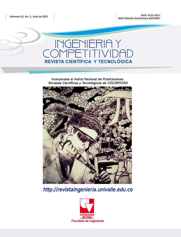
Diagnóstico de SARS-CoV-2 y métodos alternativos innovadores basados en fibra óptica.
Palabras clave:
Biosensor, COVID-19, Diagnóstico clínico, Medicina, Tecnología y Fibra óptica.Contenido principal del artículo
Este trabajo muestra una revisión mundial de los métodos diagnósticos del SARS-CoV-2, analizando sus efectividades y sensibilidades. Con un énfasis especial en los biosensores, particularmente, los que se basan en la tecnología de las fibras ópticas, explicando de manera simple su funcionamiento y su capacidad para detectar enfermedades como el SARS-CoV-2. Con estos avances tecnológicos, el diagnóstico clínico será: rápido, económico y podrá aplicarse en pacientes que vivan en lugares remotos donde no hay hospitales ni laboratorios clínicos, debido a la pobreza, las dificultades geográficas o violencia que son factores que se encuentran en Colombia.
(1) WHO and others. Protocol: Real-time RT-PCR assays for the detection of SARS-CoV-2 Institut Pasteur, Paris. Geneva: World Health Organization. 2020. Available from: https://www.who.int/docs/default-source/coronaviruse/real-time-rt-pcr-assays-for-the-detection-of-sars-cov-2-institut-pasteur-paris.pdf?sfvrsn=3662fcb6_2.
(2) Huang JC, Chang Y-F, Chen K-H, Su L-C, Lee C-W, Chen C-C, et al. Detection of severe acute respiratory syndrome (SARS) coronavirus nucleocapsid protein in human serum using a localized surface plasmon coupled fluorescence fiber-optic biosensor. Biosensors and Bioelectronics.2009;25(2):320–5. https://doi.org/10.1016/j.bios.2009.07.012.
(3) Weinstein MC, Freedberg KA, Hyle EP, Paltiel AD. Waiting for Certainty on Covid-19 Antibody Tests—At What Cost? N Engl J Med. 2020;383:e37. https://doi.org/10.1056/NEJMp2017739.
(4) Woloshin S, Patel N, Kesselheim AS. False Negative Tests for SARS-CoV-2 Infection—Challenges and Implications. N Engl J Med. 2020;383:e37. https://doi.org/10.1056/NEJMp2015897.
(5) Sassolas A, Blum LJ, Leca-Bouvier BD. Immobilization strategies to develop enzymatic biosensors. Biotechnology advances. 2012;30(3):489–511. https://doi.org/10.1016/j.biotechadv.2011.09.003.
(6) Iniewski K, Rajan G, Krzysztof Iniewski. Optical Fiber Sensors Advanced Techniques and Applications. 1st ed. Rajan G, editor. Boca Raton (FL): CRC Press; 2017. 575 p.
(7) Candiani A, Bertucci A, Giannetti S, Konstantaki M, Manicardi A, Pissadakis S, et al. Label-free DNA biosensor based on a peptide nucleic acid-functionalized microstructured optical fiber-Bragg grating. Journal of biomedical optics. 2013;18(5):057004.https://doi.org/10.1117/1.JBO.18.5.057004.
(8) Gonçalves HM, Moreira L, Pereira L, Jorge P, Gouveia C, Martins-Lopes P, et al. Biosensor for label-free DNA quantification based on functionalized LPGs. Biosensors and Bioelectronics. 2016;84:30–6. https://doi.org/10.1016/j.bios.2015.10.001.
(9) Del Villar I, Zamarreño C, Hernaez M, Sanchez P, Arregui F, Matias I. Generation of surface plasmon resonance and lossy mode resonance by thermal treatment of ITO thin-films. Optics & Laser Technology. 2015;69:1–7. https://doi.org/10.1016/j.optlastec.2014.12.012.
(10) Del Villar I, Zamarreño CR, Hernaez M, Arregui FJ, Matias IR. Lossy mode resonance generation with indium-tin-oxide-coated optical fibers for sensing applications. Journal of Lightwave Technology. 2010;28(1):111–7. https://doi.org/10.1109/JLT.2009.2036580.
(11) Maya YC, Villar I Del, Socorro AB, Corres JM, Botero-Cadavid JF. Optical Fiber Immunosensors Optimized with Cladding Etching and ITO Nanodeposition. In: 2018 IEEE Photonics Conference (IPC). Reston, VA, USA: IEEE; 2018. p. 1–2. https://doi.org/10.1109/IPCon.2018.8527306.
(12) Rijal K, Leung A, Shankar PM, Mutharasan R. Detection of pathogen Escherichia coli O157: H7 AT 70 cells/mL using antibody-immobilized biconical tapered fiber sensors. Biosensors and Bioelectronics. 2005;21(6):871–80. https://doi.org/10.1016/j.bios.2005.02.006.
(13) Chiavaioli F, Trono C, Giannetti A, Brenci M, Baldini F. Characterisation of a label-free biosensor based on long period grating. Journal of biophotonics. 2014;7(5):312–22. https://doi.org/10.1002/jbio.201200135.
(14) Dudley R, Edwards P, Ekins R, Finney D, McKenzie I, Raab G, et al. Guidelines for immunoassay data processing. Clinical chemistry.1985;31(8):1264–71.https://doi.org/10.1093/clinchem/31.8.1264.
(15) Stefan Melanie I., Novère NL. Cooperative binding. PLOS Computational Biology. 2013 Jun;9(6):e1003106. https://doi.org/10.1371/journal.pcbi.1003106.
(16) HILL AV. The possible effects of the aggregation of the molecules of haemoglobin on its dissociation curves. J. Physiol. 1910;40:4–7. Available from: https://ci.nii.ac.jp/naid/10020096935/en/.
(17) Wang C, Li W, Drabek D, Okba NM, Haperen R van, Osterhaus AD, et al. A human monoclonal antibody blocking SARS-CoV-2 infection. Nature communications. 2020;11(1):2251. https://doi.org/10.1038/s41467-020-16256-y.
(18) Matushek SM, Beavis KG, Abeleda A, Bethel C, Hunt C, Gillen S, et al. Evaluation of the EUROIMMUN Anti-SARS-CoV-2 ELISA Assay for detection of IgA and IgG antibodies. Journal of Clinical Virology. 2020;129:104468. https://doi.org/10.1016/j.jcv.2020.104468
(19) Nguyen T, Duong Bang D, Wolff A. 2019 novel coronavirus disease (COVID-19): paving the road for rapid detection and point-of-care diagnostics. Micromachines. 2020;11(3):306. https://doi.org/10.3390/mi11030306.
(20) Kakodkar P, Kaka N, Baig M. A comprehensive literature review on the clinical presentation, and management of the pandemic coronavirus disease 2019 (COVID-19). Cureus. 2020;12(4):e7560. https://dx.doi.org/10.7759%2Fcureus.7560.
(21) Bachelet VC. ¿Conocemos las propiedades diagnósticas de las pruebas usadas en COVID-19? Una revisión rápida de la literatura recientemente publicada. Medwave. 2020;20(3):e7891. https://doi.org/10.5867/medwave.2020.03.7891.
(22) Cai X, Chen J, Hu J, Long Q, Deng H, Fan K, et al. A Peptide-based Magnetic Chemiluminescence Enzyme Immunoassay for Serological Diagnosis of Corona Virus Disease 2019 (COVID-19). The Journal of Infectious Diseases. 2020;222(2):189-93. https://doi.org/10.1093/infdis/jiaa243.
(23) Carter LJ, Garner LV, Smoot JW, Li Y, Zhou Q, Saveson CJ, et al. Assay techniques and test development for COVID-19 diagnosis. ACS Cent. Sci. 2020;6(5):591–605. https://doi.org/10.1021/acscentsci.0c00501.
(24) Chen X, Tang Y, Mo Y, Li S, Lin D, Yang Z, et al. A diagnostic model for coronavirus disease 2019 (COVID-19) based on radiological semantic and clinical features: a multi-center study. European radiology. 2020;30:4893–902. https://doi.org/10.1007/s00330-020-06829-2.
(25) Guo L, Lili R, Siyuan Y, Meng X, Chang D, Fan Y, et al. Profiling early humoral response to diagnose novel coronavirus disease (COVID-19). Clinical Infectious Diseases. 2020 Jul 28;71(15):778-85. https://doi.org/10.1093/cid/ciaa31
Downloads

Esta obra está bajo una licencia internacional Creative Commons Atribución-NoComercial-CompartirIgual 4.0.
Los autores que publican en esta revista están de acuerdo con los siguientes términos:
Los autores ceden los derechos patrimoniales a la revista y a la Universidad del Valle sobre los manuscritos aceptados, pero podrán hacer los reusos que consideren pertinentes por motivos profesionales, educativos, académicos o científicos, de acuerdo con los términos de la licencia que otorga la revista a todos sus artículos.
Los artículos serán publicados bajo la licencia Creative Commons 4.0 BY-NC-SA (de atribución, no comercial, sin obras derivadas).

